Monday 18 November 2013

Paper review

The Paper I decided to review can be seen through this link. http://onlinelibrary.wiley.com/doi/10.1111/j.1365-2303.2011.00901.x/pdf

Title: Sclerosing angiomatoid nodular transformation (SANT) of spleen: a case report describing cytology, histology, immunoprofile and differential diagnosis
* Not mentioned in paper
Background:
                There are various conditions were a lesion like formation forms on the spleen. Our abilities to determine what these lesions are while the spleen is still within the patient are limited. They can mimic malignant tumors which may result in a splenectomy (removal of the spleen, it is not a vital organ). Sclerosing angiomatoid nodular transformation (SANT) is a lesion which affects the spleen but is believed to be a reactive condition oppose to the neoplasm.


Introduction:
                The paper looks at a spleen taken from a 48 year old man who was experiencing pain in the pelvic region, towards the back. Computed tomography scans showed a vascular mass within the spleen and the spleen was removed because of the risk of radiological suspicion of angiosarcoma. There was no histological or cytological sampling done before the surgery. The authors of the paper are trying to determine some histopathology, cytology and immunohistochemical features of SANT as there has not been any real report of these test done in literature so far.


Materials and methods
                The spleen from the 48 year old man who recently undergone a splenectormy was measure and examined. Cytological smears were carried out by scraping the lesion and smearing onto glass slides. The slides were fixed in 96% alcohol or air dried. They were then stained with papanicolaou (*A series of dyes used to give colors to specific things) and May-grunwald-giemsa (*Stain negative molecules purples) methods.
                The spleen was held in 10% formaldehyde, tissue blocks were paraffin embedded and 5 micrometer sections were cut and stained with haematoxylin and eosin. Immunocytochemical stains for CD34, CD31, CD8, CD23(* Dyes based on specfic antigen which differ in cells types, allows the differeniation of cell types on slide)  and anaplastic lymphoma kinase(*Tests for a common cancer causing kinase) were performed.  Also Ziehl-Neelsen and warthin-starry silver stain were carried out for possible microrganisms, as well as Epstein-Barr virus status was investigated by in situ hybridization.

Results:
                Cytologically: there were several differences from normal spleen tissues, Clusters of irregulars stromal figures (angiomatoid nodules).Composed of fibrous stroma and dispersed stromal cells with oval-spindle nuclei and scanty cytoplasm.


 Some had a round nuclei with more cytoplasm. This was shown to have haemosiderin pigment. 


Capillaries transpassing some of these stromal nodules.


                There was some convoluted nuclear outlines, nuclear grooves and small nucleoli. 


There was no cytological atypia cells, mitotic figure, giant cells or necrosis.


The legion was characterized vascular nodules and slit like spaces. The area did show many extravasated red blood cells and haemosidern pigment was plentiful.  The indivual nodules were seperated inflammatory cells. Shown below.

The different types of endothelial cells of vascular cells highlighted by the used of the immunohistochemmical panel. Capillaries( CD31+  CD34+ CD8-) Sinusoid type spaces (CD31+ CD34- CD+_ and small vien (CD31+ CD34 - CD8-) Shown respectively below.


No CD23 or ALK kept stain, no microorganisms were found or viral through EBER.

Discussion:

          The discussion compares the SANT cytology and histology to certain cancer masses that could be present in the spleen. Believed to resemble granulomatous inflammation. The vascular characteristics and lack of certain granuloma features seem to be the best way to diagnose this disorder. The cause for these formation appears to be unknown.

Critique

        The paper showed some finding to help further diagnose SANT. The introduction was particularly short, with many things that could of been mentioned in the introduction being explained in the discussion which seem counter intuitive. Other then that the paper was short and directly on the topic which made it an easy read. 
        The picture of the tissue samples were well clear pictures. However it would be much better with a control picture of normal spleen tissue to compare it to. Also a picture of a normal spleen compared to a SANT spleen without magnification to see the mass form lesion. Comparing the cancer like masses in the discussion would have been a better way to visualize the differences.

    The paper was also fairly short, I believe this was due to the nature of the disease. The experiments needed to be done on a "fresh" spleen and due to the rarity of the disease I assume this is why it only was completed on one spleen. Future experiments on other cases could provide a better paper were it summarizes the findings in several papers.

   Also had I had some trouble with the language in the paper, a better introduction could help with that although the authors probably intended this to be viewed by people with a stronger back ground then I. 


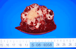
Here is a spleen picture with SANT. Not from paper


Thursday 24 October 2013

The Spleen


Introduction
The spleen is a relatively small organ which is located behind the stomach (see figure above) and is usually around 5 inches wide. It can be divided into two sections; the red pulp and the white pulp. The red pulp is used to filter blood and the white pulp is used for antigen detection and elimination.


 The spleen itself contains a lot of vessel tissues and can be used to store significant amounts of blood which is useful to help counter act loss of blood trauma (Cesta 2006).This allows a large amount of blood flow to the organ which will help it complete its already mentioned functions.  This blog will discuss the tissues in the spleen which allow it to be such a spleen-did organ.


General Structure
The spleen can be divided into several sections.

Capsule: Is the outer layer of the spleen. It is mostly dense fibrous, elastic fibres and smooth muscle.


Trabeculae: Smooth muscle and fibroelastic tissue that spread from the capsule inward in an irregular shape. Trabeculae also contain blood and lymph vessels and nerves. Aids in structure, helps with movement of blood to the parts of the spleen.


Red Pulp
The red pulp is used to filter the blood removing old red blood cells and reuse their valuable iron for production of new erythrocytes and other metabolic processes.  This is accomplished when the blood enters the cords(shown in the picture below).
Cords: The structure is most fibroblasts and reticular fibres which house a lot of macrophages. Macrophages here are what breaks down the old red blood cells. The cords act as an open circulatory system which lack endothelial lining (Mebius and Kraal 2005)


Venous sinuses penetrate the cords and regulate the passage of erythrocytes into them and thus back into the blood. These vessels have a specialised structure which is shown below. It involves endothelial cells that have stress fibres connecting throughout them parallel. Contraction of the stress fibres creates small slits that allow the red blood cells to enter the venous sinuses (Mebius and Kraal 2005). Older red blood cells have more rigid cell membranes and have more trouble moving through these slits. This leaves them in the cords with the large amount of macrophages.  The red blood cells are targeted by the macrophages and are ingested in phagolysosomes. This will hydrolyse the cell and destroy the hemoglobin proteins releasing Iron.  Iron can be a dangerous molecule in biological systems, and is kept in proteins called ferritin, or in larger quantities the transporter protein transferrin(Mebius and Kraal 2005).


Haemolytic bacteria can pose a big threat here in the spleen. Haemolytic bacteria break down red blood cells for iron and other nutrients, so the spleen has ways to deal with these bacteria. Many of these bacteria for example will secrete molecules known as siderophores which bind iron and using specific transport proteins on the bacteria membrane will take in the siderophore (which have now have iron). The macrophages of the spleen can be easily induced to secrete the protein lipocalin-2 which binds the siderophores and will prevent bacteria from taking the iron.(Ratledge and Dover 2000)


White Pulp
The figure below shows the white pulp organisation in which you have a central arteriole enter the white pulp nodule which is composed of B cell zone(also called follicle), marginal zone and the T-cell zone(sometimes referred to as the PALS which stands for periarteriolar lymphoid sheath).  This actually is a very similar arrangement as the lymph node(Cesta 2006).


A little reminder of the function of immune cells!
T-cells are immune cells that come in several types.  T helper cells which help other cells recognise, mature or activate. Cytotoxic T-cells which kill “bad” host cells which will be recognise for cancer patterns, intracellular bacteria or viruses. (Other sometimes cause problems with autoimmune problems,  transplant rejections etc). Regulatory T cells which help control immune response to desired levels. Natural killer t-cells which help the innate response trigger the adaptive responses of the immune system. Rely on cell surface receptors of other immune cells to activate.


B Cells are function to produce specific antibodies which will bind to antigens. Binding to antigen allow them to be identified by other immune processes and to cause agglutination. (Basically bridge a large amount of antigens together to make an insoluble clump which prevent antigen from doing certain things and allow easy identification.)  B cells often come from clones and antigen specific recognition needs to be effective.
The follicle and T-cell are kept their organisation by specific chemokines. The T-cells can interact with dendritic cells and passing B-cells. The follicles are where clonal selection takes place for B-cells. The bulk of blood travels through the white pulp upon entering the spleen. This allows a good way to detect any pathogens or foreign material that may be in the blood.

 Marginal Zone

This is a layer which separates the white and red pulp, which is argued to be considered part of the white pulp or its own layer which divides the red and white pulps. Its purpose is to screen the any viruses or bacteria. There is a band of macrophages which separate the white pulp and the marginal zone which are called as marginal zone metallophilic macrophages characterised by their adhesion molecules. The marginal zone itself has layers of reticular fibroblasts where more immune cells reside including specialised B cells which can be differentiate by the types of antibodies the produced compared to the follicle region.(Mebius and Kraal 2005)


Pathology
The most common problem with the spleen is spleen is called splenomegaly which is easily noticed by enlargement of the spleen. This caused by a variety of conditions such as congestion, neoplasm, injection, hemolytic anemia, extramedullary hematoposis or trauma. 


The genetic disorder Gaucher disease causes spleenic enlargement as well. This disease is categorised by macrophages carrying too much glycolipids. This disease effects many organs including the spleen. Shown below (Cox et al., 1997)

Cystic lymphangiomatosis is another rare disease which again effects several organs and is caused by the a development issue with lymphatic tissues which can again effect the spleen (Mohanana et al., 2009)


References
Cesta. M (2006) Normal structure, function and histology of the spleen. Toxicol Pathol 34:455-466

Ratledge C, Dover L (2000) Iron metabolism in pathogenic bacteria Annu. Rev.Microbiology  54: 881-941

Mebius R, Kraal 2005. Structure and Function of the spleen. Nature Vol 5 606-616

Wright, Dennis Wilkins, Bridget , Illustrated Pathology of the Spleen 03/2000             

Cox T, Shcofield J, 1997. Gaucher's disease: Clinical features and natural historyBailli~re's Clinical Haematology-- 657 Vol. 10, No. 4, December 1997


S. Mohana, N.V. Seethalekshmy, K. Pavithran: Splenic Cystic lymphangiomatosis presenting with massive splenomegaly. The Internet Journal of Pathology. 2009 Volume 8 Number 1. DOI: 10.5580/21ca


 http://instruction.cvhs.okstate.edu/histology/HistologyReference/hrlym.htm  
 http://www.deltagen.com/target/histologyatlas/atlas_files/hematopoietic/spleen_red_pulp_40x.htm
 http://www.cell.com/immunity/image_resource-spleen
http://archive.ispub.com/journal/the-internet-journal-of-pathology/volume-8-number-1/splenic-cystic-lymphangiomatosis-presenting-with-massive-splenomegaly.html#sthash.PKWmivin.dpbs