The Paper I decided to review can be seen through this link. http://onlinelibrary.wiley.com/doi/10.1111/j.1365-2303.2011.00901.x/pdf
Title: Sclerosing angiomatoid nodular transformation (SANT) of spleen: a case report describing cytology, histology, immunoprofile and differential diagnosis
* Not mentioned in paper
Background:
There
are various conditions were a lesion like formation forms on the spleen. Our
abilities to determine what these lesions are while the spleen is still within
the patient are limited. They can mimic malignant tumors which may result
in a splenectomy (removal of the spleen, it is not a vital organ). Sclerosing
angiomatoid nodular transformation (SANT) is a lesion which affects the spleen
but is believed to be a reactive condition oppose to the neoplasm.
Introduction:
The
paper looks at a spleen taken from a 48 year old man who was experiencing pain
in the pelvic region, towards the back. Computed tomography scans showed a vascular
mass within the spleen and the spleen was removed because of the risk of
radiological suspicion of angiosarcoma. There was no histological or
cytological sampling done before the surgery. The authors of the paper are
trying to determine some histopathology, cytology and immunohistochemical
features of SANT as there has not been any real report of these test done in literature so
far.
Materials
and methods
The
spleen from the 48 year old man who recently undergone a splenectormy was
measure and examined. Cytological smears were carried out by scraping the
lesion and smearing onto glass slides. The slides were fixed in 96% alcohol or
air dried. They were then stained with papanicolaou (*A series of dyes used to give colors to specific things) and May-grunwald-giemsa (*Stain negative molecules purples) methods.
The
spleen was held in 10% formaldehyde, tissue blocks were paraffin embedded and 5
micrometer sections were cut and stained with haematoxylin and eosin.
Immunocytochemical stains for CD34, CD31, CD8, CD23(* Dyes based on specfic antigen which differ in cells types, allows the differeniation of cell types on slide) and anaplastic lymphoma
kinase(*Tests for a common cancer causing kinase) were performed. Also
Ziehl-Neelsen and warthin-starry silver stain were carried out for possible
microrganisms, as well as Epstein-Barr virus status was investigated by in situ
hybridization.
Results:
Cytologically:
there were several differences from normal spleen tissues, Clusters of irregulars stromal figures (angiomatoid nodules).Composed of fibrous stroma and dispersed stromal cells with oval-spindle nuclei and scanty cytoplasm.
Some had a round nuclei with more cytoplasm. This was shown to have haemosiderin pigment.
Capillaries transpassing some of these stromal nodules.
There was some convoluted nuclear outlines, nuclear grooves and small nucleoli.
There was no cytological atypia cells, mitotic figure, giant cells or necrosis.
The legion was characterized vascular nodules and slit like spaces. The area did show many extravasated red blood cells and haemosidern pigment was plentiful. The indivual nodules were seperated inflammatory cells. Shown below.
The different types of endothelial cells of vascular cells highlighted by the used of the immunohistochemmical panel. Capillaries( CD31+ CD34+ CD8-) Sinusoid type spaces (CD31+ CD34- CD+_ and small vien (CD31+ CD34 - CD8-) Shown respectively below.
No CD23 or ALK kept stain, no microorganisms were found or viral through EBER.
Discussion:
The discussion compares the SANT cytology and histology to certain cancer masses that could be present in the spleen. Believed to resemble granulomatous inflammation. The vascular characteristics and lack of certain granuloma features seem to be the best way to diagnose this disorder. The cause for these formation appears to be unknown.
Critique
The paper showed some finding to help further diagnose SANT. The introduction was particularly short, with many things that could of been mentioned in the introduction being explained in the discussion which seem counter intuitive. Other then that the paper was short and directly on the topic which made it an easy read.
The picture of the tissue samples were well clear pictures. However it would be much better with a control picture of normal spleen tissue to compare it to. Also a picture of a normal spleen compared to a SANT spleen without magnification to see the mass form lesion. Comparing the cancer like masses in the discussion would have been a better way to visualize the differences.
The paper was also fairly short, I believe this was due to the nature of the disease. The experiments needed to be done on a "fresh" spleen and due to the rarity of the disease I assume this is why it only was completed on one spleen. Future experiments on other cases could provide a better paper were it summarizes the findings in several papers.
Also had I had some trouble with the language in the paper, a better introduction could help with that although the authors probably intended this to be viewed by people with a stronger back ground then I.
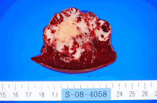
Here is a spleen picture with SANT. Not from paper


No comments:
Post a Comment